Project
PBPK modeling
Scientific modeling work in physiologically-based pharmacokinetics (PBPK) for intravenously delivered nanoparticle drugs: model structure design, distribution equations, parameter estimation, sensitivity analysis, and validation.
Context
PBPK modeling uses compartments that match real organs and blood flow to predict where a drug ends up in the body. For nanoparticle drugs delivered intravenously, this matters because you cannot easily sample every tissue, and formulation choices, including whether the particle dissolves, how big it is, and whether immune cells capture it, change the governing equations. This project covers model structure design, the corresponding mathematics, parameter estimation, sensitivity analysis, and validation against animal data.
What I worked on
Whole-body PBPK structures for nanoparticle carriers such as colloidal gold, liposomes, and nanocrystals; deciding when to use flow-limited versus membrane-limited assumptions; adding a macrophage uptake subcompartment for colloidal particles that do not dissolve; estimating parameters and running sensitivity analyses; and checking predictions against measured biodistribution.
Tutorial paper
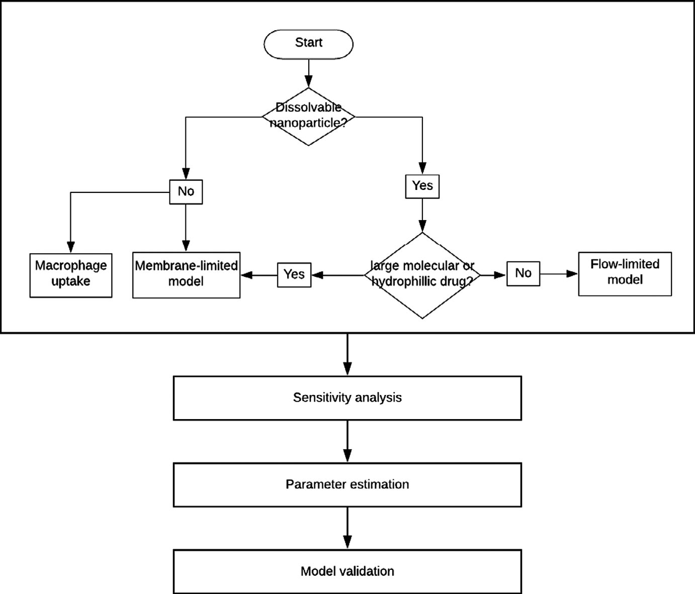
I wrote a first-author tutorial in CPT: Pharmacometrics & Systems Pharmacology (2022) that walks through building a PBPK model for IV-delivered nanoparticle drugs. The core idea is to answer a few binary questions about the formulation (does it dissolve? is it membrane- or flow-limited? is macrophage uptake expected?) and then let the answers select the model structure before any equations are written. The paper covers the decision flow, four canonical compartmental layouts, the mass-balance ODE framework, subcompartmentalization, parameter fitting, sensitivity testing, and validation. It is open-access and meant for students and people entering the field.
Tools and methods
Compartmental and whole-body PBPK modeling, systems of ODEs for mass balance across organ compartments (flow-limited and membrane-limited formulations), nanoparticle-specific terms for dissolution and macrophage absorption/desorption, parameter estimation, sensitivity analysis, and validation against in vivo biodistribution using R² of log-transformed predicted versus measured concentrations.
What I learned
A model is most useful when its limitations are explicit. For nanoparticle drugs, whether the formulation is dissolvable or colloidal, large/hydrophilic or small/lipophilic, and with or without phagocyte uptake almost entirely determines the right model structure. Getting that decision wrong upstream wastes every fitting and sensitivity step that follows.
Status and next questions
Continuing interest in modeling questions that connect nanoparticle behavior to regulatory decision-making, particularly where preclinical biodistribution data have to support dose justification.
Technical detail from the tutorial
The figures and walkthrough below are drawn from the open-access tutorial paper and reproduced here under its Creative Commons BY-NC license. They cover the decision flow for selecting a PBPK model structure, the resulting whole-body compartmental layouts, and the macrophage subcompartment used for colloidal particles.
Figures and equations adapted from: Le A-D, Wearing HJ, Li D. Streamlining physiologically-based pharmacokinetic model design for intravenous delivery of nanoparticle drugs. CPT Pharmacomet Syst Pharmacol. 2022;11:409–424 [1]. Open-access, CC BY-NC.
Pick the structure from the physicochemistry, not the other way around
The tutorial approaches model design as a short chain of yes-or-no questions about the drug formulation. Does the nanoparticle dissolve in aqueous fluid, or does it stay intact as a colloid? Is it large and hydrophilic, so crossing membranes is the slow step, or small and lipophilic, so blood flow is the bottleneck? Should macrophage uptake be expected? The answers pick between a perfusion-limited (flow-limited) and a permeability-limited (membrane-limited) layout, and decide whether dissolution and macrophage subcompartments are needed [1]. Answering these questions before writing equations keeps the later parameter fitting and validation grounded.
Four canonical whole-body structures
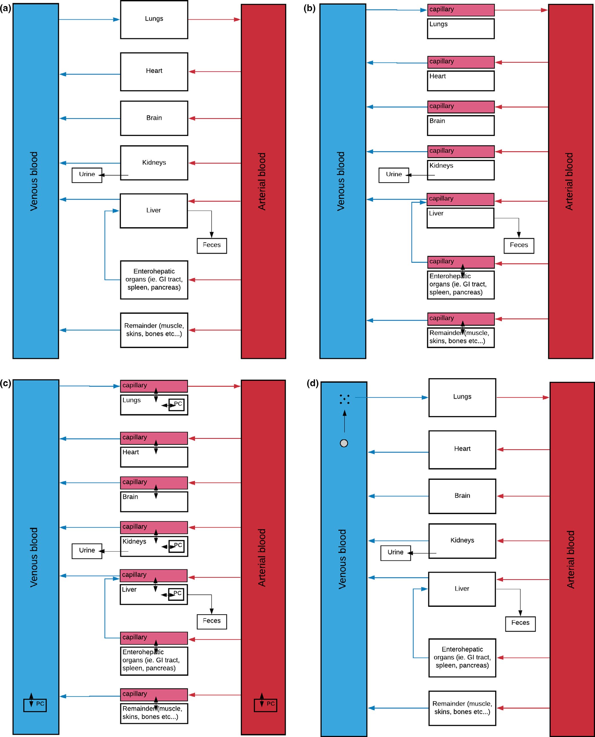
The paper illustrates four layouts that come out of the decision flow. In (a), a conventional non-nanoparticle drug uses a flow-limited structure: each tissue is a single well-mixed compartment fed by arterial blood and drained to venous blood. In (b), a colloidal nanoparticle that does not dissolve uses a membrane-limited structure, splitting each tissue into a capillary and a tissue subcompartment separated by a permeability term. In (c), the same membrane-limited layout adds phagocytizing-cell (PC) subcompartments inside tissues with high macrophage density. In (d), a dissolvable nanoparticle uses a flow-limited structure where particles release free drug at rate k_rel into the surrounding compartment [1].
Mathematical framework
Each tissue compartment is described by a mass-balance ODE built from the law of mass action. For a baseline flow-limited tissue, compartment 2 fed by compartment 1 obeys Eq. (1), where Q is blood flow, C₁ and C₂ are concentrations, and k_p is the partition coefficient (a distribution coefficient for colloidal nanoparticles). Tissues that clear drug add an excretion term dM_ex/dt collecting renal and biliary clearance, giving Eq. (2). For colloidal nanoparticles, a permeability factor X/(1+X) gates flow across the capillary–tissue membrane, giving Eq. (5). For dissolvable nanoparticles, a first-order release term k_rel · V_t · C couples the particle and dissolved-drug equations with opposite signs on each side, giving Eq. (4) [1].
Macrophage uptake as a physiological subcompartment
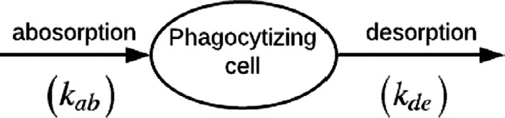
Because colloidal nanoparticles do not dissolve, they are subject to immune clearance. The tutorial treats macrophage uptake as a small two-step subcompartment with absorption rate k_ab and desorption rate k_de, placed inside tissues with high macrophage density such as the liver (Kupffer cells). The governing equation for those tissues picks up an extra term, (k_de · M_{2,m}) − (k_ab · V_2 · C_2), where M_{2,m} is the amount currently held in macrophages, giving Eq. (6). This term is what allows the model to capture the long tissue residence times that colloidal nanoparticles exhibit in vivo [1].
Parameters, sensitivity, and validation
Inputs split into three groups. Physicochemical properties, such as partition coefficient, molecular weight, and solubility, depend only on the drug. Drug–biological properties, such as fraction unbound, Michaelis–Menten constants, and permeability, depend on both the drug and the organism. Anatomic and physiological properties, such as organ volumes, surface areas, blood flows, and expression levels, come from the organism. Many are taken from literature or databases; the rest are fitted to in vivo biodistribution data. Nanoparticle-specific additions include the release constant, maximum uptake rate in phagocytic cells, Hill coefficient, and desorption constant. Sensitivity analysis helps decide which parameters are worth the effort to estimate. For instance, whether the dissolution rate of a protein-corona-coated nanoparticle actually moves the output. Validation is performed on held-out data, usually by comparing log₁₀(predicted) against log₁₀(measured) tissue concentrations via R² [1].
References
- [1]Le A-D, Wearing HJ, Li D. Streamlining physiologically-based pharmacokinetic model design for intravenous delivery of nanoparticle drugs. CPT: Pharmacometrics & Systems Pharmacology. 2022;11(4):409–424. link